The brain, the most complex organ in our body, has numerous functions: it regulates the functions of many organs within our body, allows us to control our movements, and manages our thoughts, memory, and speech. Your brain constantly absorbs information from your surroundings every day and learns from it, through numerous chemical reactions that take place inside the cells in your head. The images, feelings, and experiences are all formed by your brain.
1. What happens in the brain?
The brain is composed of billions of cells called neurons and synapses which are interconnected forming a vast network. Each neuron is composed of a body and dendrites enabling information to travel to and from the brain. When the brain reacts, these neurons release chemicals called neurotransmitters to neighboring neurons. In the neuronal connections, electrical signals travel along axons triggering the changes in chemical balance in the brain and, as a result, create an electrical field. These changes, often referred to as potentials, don’t last long because the brain continues creating information that causes these potentials to change continuously. The sensors in the EEG machines quickly pick up these electric fields in a very precise manner which are later used by amplifying these recordings.
2. What is EEG and its principles?
EEG stands for electroencephalography which is a monitoring method that records the electrical activity of the brain. Electrode sensors are placed on the scalp and amplify the small voltage changes that arise from electrical activity in the brain. EEG has been a significant tool for research and diagnostic tests in that it allows us to understand how our brains operate and react under certain conditions or symptoms. EEG provides excellent time resolution, allowing you to detect activity within cortical areas even in the scale of milliseconds. One of the biggest advantages to EEG is that it is one of the fastest and cheapest imaging techniques available and often has a high sampling rate.
The number of EEG sensors vary among devices, but the more sensors used, more regional data can be obtained. The 10-20 system is an international method that standardizes the placement of the electrodes, or sensors, on the individual’s scalp.
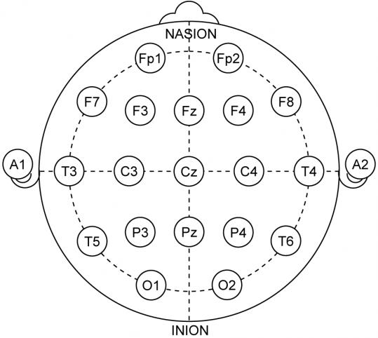
For better understanding, the image below illustrates how this method works. It demonstrates where the electrodes should be placed, where regions A1 and A2 are the left and right ears respectively, the nasion as the point between the eyes and the inion as the back of a head. Each placement of the electrodes is at a 10% or 20% distance of the front-back or left-right distance of a person’s head.

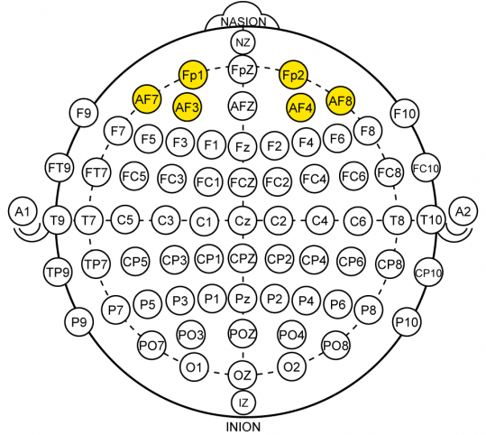
As for the placement of the 10-10 system, divide all the 20% distance regions of the 10-20 system by half, and you will be able to see the regions that are used for the EEG electrode placement.
3. Lobes of the Brain: Structure and Functions
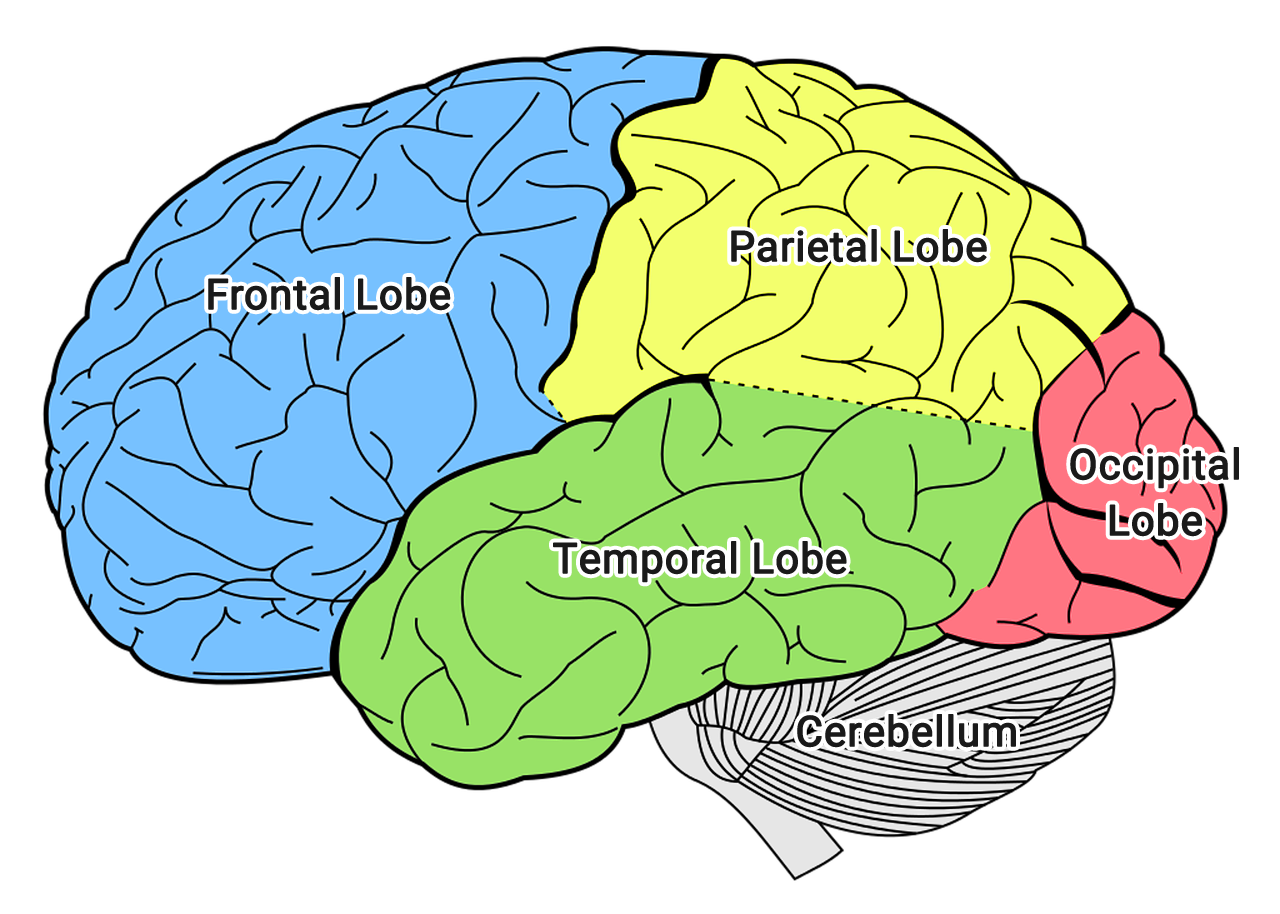
The outermost layer of the brain is called the cerebral cortex. Both cerebral hemispheres are further divided into lobes. Here are each of the lobes with a brief description of their functions:
- Frontal lobe – located in the front of the brain. The region is responsible for cognitive skills such as solving problems, planning, making decisions, as well as controlling emotions and our behavior (executive functions).
- Temporal lobe – located on the side of the brain. The region is responsible not only for primary auditory perception in terms of processing and understanding sounds like hearing but also language processing and speech production (left temporal cortex including Broca’s and Wernicke’s areas). The region also plays a role of long-term memory (deep temporal structures including the hippocampus) and managing complex visual information including faces.
- Parietal lobe – located above the temporal lobe and right behind the frontal lobes. The region is responsible for sensory and motor functions of the body. The parietal lobe is also in relation to the skills such as hand-eye coordination (visuospatial processing).
- Occipital lobe – located at the back of the brain. The region plays an important role in vision, in terms of processing all the visual information such as depth, distance, and location of the objects (orientation, spatial frequency).
When an individual undergoes certain types of activities such as learning, exercising, and memorizing, the regions that are associated with those activities will activate. Detecting the sub-second changes of EEG signals can give you insight on what areas of the brain are processing information at a given time. However, it is important to know that the measurements given by EEGs cannot diagnose disorders themselves but allow us to have a clear overview of confirming or ruling out various conditions including seizure disorders and head injury.
Why is the prefrontal cortex important?
The prefrontal cortex is located at the front part of the frontal lobe. The prefrontal cortex is implicated in planning complex cognitive behavior, personality expression, decision making, and moderating social behavior. At Looxid Labs, we focus on the prefrontal cortex because we are aiming to provide a compatible interface for virtual reality (VR) and comfortability in terms of usability of VR headsets. Many headsets will need support mechanisms to hold the VR devices which impede the placement of sensors on some regions of the scalp. By placing the EEG sensors in an area that does not obstruct the functionality of a head mounted display, the most logical area to place these electrodes is the forehead area, which corresponds to the prefrontal cortex of the brain.
4. Different Types of Brainwaves
When your brain is in a certain state of mind, the frequency patterns of the brainwave changes, giving insight into the cognitive process. The frequency patterns of brainwaves are categorized into 5 different types, each of which suggests different states of the mind. Previous neuroscience studies confirm that each brainwave type is crucial to all aspects of brain functioning such as what is really undergoing in our minds.
- Delta Waves (0.5Hz ~ 4Hz)
The lowest frequency of all brainwaves, delta waves are prominent when an individual is in deep stages of sleep, more specifically during the non-REM sleep. When these waves are the strongest, one can infer that the individual is going through good rest, consequently allowing the brain to process learned information and skills. Abnormalities in the presence of this wave frequency means that your body will not be able to produce the hormones that promote healing and regeneration of the body. When it comes to sleeping disorders, these are the waves to search for.

- Theta Waves (4Hz ~ 7Hz)
Theta waves appear frequently in younger children and in adults. These are indicators of drowsiness or meditative states. Also, these waves appear before an individual is about to receive new information, meaning that theta waves have a role in focused attention and processing of information, as theta waves are noticeable for more difficult tasks. The general consensus is that these waves are related with working memory and are present during REM sleep, which is when the brain is organizing and learning information.
- Alpha Waves (8Hz ~ 15Hz)
Alpha waves are present when an individual is in a relaxed state of mind. In other words, high levels of alpha waves are an indication of deep relaxation. When undergoing intense cognitive situations, these levels decline and one can induce these brainwaves by closing their eyes. Studies suggest that when alpha wave levels decrease, one is preparing themselves to pick up information or is thinking. Alpha waves aid in increasing levels of imagination, visualization, learning, and concentration.

- Beta Waves (16Hz ~ 31Hz)
Beta waves are indicators of active thinking. When an individual is alert, attentive, or awake, beta waves will be present. In particular, these oscillations correlate with execution of bodily movements. Studies have also shown that certain beta waves are indicators of an anxious or busy mind. In other words, this means that an increase of beta waves can determine that an individual is performing some kind of mental activity. The more complex mental activity required, such as critical thinking and reasoning, the more beta waves will be present.

- Gamma Waves (31Hz ~ 100Hz)
Unlike other brainwave frequencies known to be indicators of a specific state, gamma waves haven’t quite reached the same consensus as other wave forms. However, it is suggested that the presence of gamma waves indicates simultaneous processing undergoing in the brain. The presence of gamma waves during wakefulness compared to during sleep suggests that it might be a link for different stages of conscious awareness. Other studies speculate that gamma waves have control over consciousness, perception, other virtues such as altruism, and spiritual development.

5. Obtainable information using Looxid Link
As stated in the previous section, each wave frequency indicates different conditions in the brain. Likewise, depending on what you are specifically looking for, a combination of frequencies might be necessary. Our brains react to certain situations in forms that we sometimes cannot convey, which is why physiological signals like brainwaves can be a very useful tool to determine an individual’s state of mind. You can determine if a person is attentive to a university lecture, if they are stressed because of family problems, or if they are relaxed by watching a good movie. It is important to notice that not one brainwave frequency equals a certain state of mind but are merely indicators. EEG does not read your emotions or feelings; it makes sense of what one is going through.
If you try the VR mind care solution which comes along with Looxid Link, you will notice our sensors pick up your brainwave signals and quantify your frequencies in real time so that you can monitor your mental state during the session. This helps users understand how their brains react and as a result, contribute to their wellness.
6. Applicable Fields
EEG is used in a variety of fields where unbiased, unconscious data are pivotal. Different factors can affect how one expresses their minds. Hence, EEG is widely used in fields where user feedback is necessary. The following are some fields where EEG is widely known to be utilized.
- Neuro-Entertainment: Have you ever watched a movie where you thought “man, it is not engaging enough” or “this is boring”? This new field uses feedback from your brain to make the content more intense or engaging. EEG signals are effective in utilizing a person’s engagement or attention levels to make a movie more appealing to the consumer or not letting a movie be more than what the consumer can handle.
- Wellness & Healthcare: Therapy is important to recover from certain trauma or issues that one cannot overcome. EEG applied to guided therapy sessions allows for monitoring of EEG signals to observe if a certain session is helpful to the user. Physiological signs provide deeper insights than self-expressed statements. The materialization of signals before and after sessions as well as the history of these records can help soothe a user’s mind. Meditation and exposure therapy are some examples of uses in the wellness area. In addition, abnormal EEG signals of the user can help detect certain conditions before they are fatal.
- Neuromarketing: Consumer research focuses on the effectiveness of marketing campaigns. Getting to understand what catches customers’ attention and acknowledging the ways in which people perceive a marketing strategy can save a company unnecessary expenses from facts performed by neuromarketing. Self-reporting may not give deep insights, hence, EEG data can be a more resourceful option.
- Education & Training: Imagine going to class and having a lecture where the professor is able to effectively teach you, customizing the material to be more engaging, and actually being able to understand everything he or she is talking about. EEG in education is widely used in this manner, where EGG recordings during classes allow teachers to obtain reports of students’ engagement levels. As in training, companies and institutions can use EEG along with VR to allow trainees to experience real situations and get effective feedback from their mistakes in the training sessions. It allows them to be better prepared for real life situations by knowing what to expect.
References
- Cantero, JL. et al. (2004). “Gamma EEG dynamics in neocortex and hippocampus during human wakefulness and sleep”. NeuroImage 22 (3): 1271-1280.
doi: 10.1016/j.neuroimage.2004.03.014 - Davidson, RJ. (2004). “What does the prefrontal cortes ‘do’ in affect: perspectives on frontal EEG asymmetry research”. Biological Psychology 67(2004) 219-233.
doi:10.1016/j.biopsycho.2004.03.008 - Klimesch, W (1999). “EEG alpha and theta oscillations reflect cognitive and memory performance: a review and analysis”. Brain Research Reviews 29 (2-3):169-195.
doi: 10.1016/S0165-0173(98)00056-3 - Liu, NH et al. (2013). “Recognizing the Degree of Human Attention Using EEG Signals from Mobile Sensors”. Sensors 13(8): 10273-10286. doi: 10.3390/s130810273. PMID: 23939584
- Palmiero, M and Piccardi L. (2017). “Frontal EEG Asymmetry of Mood: A Mini-Review”. Front Behav Neurosci 2:224. doi: 10.3389/fnbeh.2017.00224
- Sander, C. (2018). “Changes in brain arousal (EEG-vigilance) after therapeutic sleep deprivation in depressive patients and healthy controls”. Scientific Reports 8:15087 DOI:10.1038/s41598-018-33228-x
- Whishaw IQ, Vanderwolf CH (1973). “Hippocampal EEG and behavior: changes in amplitude and frequency of RSA (theta rhythm) associated with spontaneous and learned movement patterns in rats and cats”. Behav Biol. 8 (4): 461–84. doi:10.1016/S0091-6773(73)80041-0.PMID 4350255. (Theta)
- Zhao, G et al. (2018). “Asymmetric hemisphere activation in tenderness: evidence from EEG signals”. Scientific Reports 8 (8029): DOI:10.1038/s41598-018-26133-w